Saturday 22 August
10:00
Open
Session I: Introduction
10:00-10:05
John R. Helliwell and Brian McMahon
Introduction and Welcome
10:05-10:20
John R. Helliwell
Update on activities of the IUCr Diffraction Data Deposition Working Group (DDDWG)
| Presentation  (1.26 MB)
![[J. R. Helliwell]](https://www.iucr.org/__data/assets/image/0003/113736/jrh.jpg)
John R Helliwell1 and Brian McMahon2
1 School of Chemistry, University of Manchester, M13 9PL, UK. Email: john.helliwell@manchester.ac.uk
2 IUCr, 5 Abbey Square, Chester CH1 2HU, UK. Email: bm@iucr.org
This workshop follows on from the 2012 Workshop on Diffraction Data Deposition in Bergen (http://www.iucr.org/resources/data/dddwg/bergen-workshop) and Working Group meetings at ECM28 (U. Warwick, UK; see http://forums.iucr.org/viewtopic.php?t=332) and the IUCr Congress (Montreal, Canada; http://forums.iucr.org/viewtopic.php?t=347). The Bergen Workshop identified the need for a thorough examination of current practice with metadata for raw diffraction data, and the possibility of using such a review to stimulate improved metadata characterization and handling in non-diffraction studies. This workshop will address both requirements.
In the wider scene 'Open Data' as a requirement of research publication is accelerating, whether it be derived, processed or raw data. Crystallography as a field compares well with other fields such as astronomy and particle physics in achieving 'open data' and each field finds raw data archiving challenging, especially the square kilometre array (SKA) in radio astronomy, since raw data is obviously the most voluminous. However, volume alone is not the greatest challenge. Stored raw data must be properly described so that its value and reliability can be assessed and understood, and individual data sets must be discoverable and reusable by other researchers, whether associated with formal publications or not. This is where metadata plays a key role.
We addressed technical options for achieving raw data archiving in Bergen and favoured flexibility in the physical location of the data sets, but with a key need for assigning DOIs to each raw data set. Interestingly, Nature magazine on 9 July9th 2015 highlighted 'the cloud' and commercial providers as being a preferred method for genomics data archiving. The change of attitude of the USA NIH, for example, where there were worries over the security of the commercial data store option, is significant.
Session II: Diffraction images - what can we get out?
10:20-11:00
Keynote: Loes Kroon-Batenburg
The need for metadata in archiving raw diffraction image data
| Presentation (4.43 MB)
![[L. Kroon-Batenburg]](https://www.iucr.org/__data/assets/image/0014/113351/loes.jpg)
Loes M.J. Kroon-Batenburga & John R. Helliwellb
aCrystal and Structural Chemistry, Bijvoet Center for Biomolecular Research, Utrecht University, The Netherlands
bSchool of Chemistry, Faculty of Engineering and Physical Sciences, University of Manchester, England.
Recently, the IUCr (International Union of Crystallography) initiated the formation of a Diffraction Data Deposition Working Group with the aim to develop standards for the representation of raw diffraction data associated with the publication of structural papers. Reports and minutes of DDDWG meetings can be found at forums.iucr.org. Archiving of raw data serves several goals: to improve the record of science, to verify the reproducibility and to allow detailed checks of scientific data, safeguarding against fraud or to allow reanalysis with future improved techniques.
In a special series of papers on "Archiving raw crystallographic data" (Terwilliger, 2014), we reported on our experience of transferring and archiving raw diffraction data and on the problems encountered with acquiring and deciphering sufficient metadata (Kroon-Batenburg & Helliwell, 2014).
To be able to process the raw data one needs information on the pixel geometry, information on pixel wise corrections applied, on beam polarization, wavelength and detector position amongst others, which are ideally contained in the image header. We will demonstrate that often one needs prior knowledge, evidently of how to read the (binary) detector format, but also on the set-up of goniometer geometries. This raises concerns with respect to long-term archiving of raw diffraction data. Care has to be taken that in the future unambiguous information is available i.e. one cannot simply "deposit the raw data" without such metadata details.
We made available a local raw X-ray diffraction images data archive at the Utrecht University (rawdata.chem.uu.nl), subsequently mirrored at the Tardis Raw Diffraction Data Archive in Australia, and since March 2015 made available through digital object identifiers (doi) at the eScholar University of Manchester Library data archive. Since 2013 approximately 150 GB of data was retrieved from our archive and some of the data sets which were reprocessed by other groups.
Kroon-Batenburg, L.M.J. & Helliwell, J.R. (2014) Acta Cryst. D70, 2502-2509.
Terwilliger, T.C. (2014). Acta Cryst. D70, 2500-2501.
11:00-11:20
Coffee
11:20-11:45
Wladek Minor
Crystallographic raw data: our plans and implementations within the NIH's Big Data to Knowledge resource
| Presentation (6.45 MB)
![[W. Minor]](https://www.iucr.org/__data/assets/image/0005/113756/minor.jpg) Wladek Minor
Wladek Minor
Department of Molecular Physiology and Biological Physics, University of Virginia, 1340 Jefferson Park Avenue, Jordan Hall, Room 4223, Charlottesville, VA 22908, USA
The NIH pilot project 'Integrated resource for reproducibility in macromolecular crystallography' will create a web-based archive of diffraction images collected from macromolecular samples around the world. The resource will enhance and sustain the macromolecular diffraction data comprising the primary data sources for macromolecular atomic coordinates in the Protein Data Bank (PDB). The project will develop tools that will extract metadata from images alone, or from a combination of information obtained from a PDB deposit and diffraction images. All of the metadata needed for automatic determination and re-determination of macromolecular structures will be collected. Currently, the project has more than 1500 data sets and a preliminary system for extracting certain types of metadata. The data mining tools developed will allow for analysis of single experiments, as well as sets of experiments performed using various synchrotron and home based sources. Diffraction sets and metadata will be available from the project's website at http://www.proteindiffraction.org, or through a link on a PDB deposits page on the RCSB PDB website. This talk will present initial results of data mining performed on the archive.
11:45-12:10
Michael E. Wall
Metadata needed for the full exploitation of diffuse scattering data from protein crystals
| Presentation (3.17 MB)
![[M. Wall]](https://www.iucr.org/__data/assets/image/0019/113491/IMG_1078.jpg)
Michael E. Wall
Los Alamos National Laboratory, CCS-3 MS B256, Los Alamos, NM 87545, USA
Technical release #: LA-UR-15-23866
I will review efforts to model motions of crystalline proteins using diffuse X-ray scattering. This work requires analysis of raw diffraction images, which are mostly inaccessible in public databases. There is an abundance of potential metadata from these studies, including information about the analysis methods, measurements of diffuse intensity, and results from the modeling. The time is now ripe for integrating diffuse scattering into traditional crystallography: modern beam lines and detectors are enabling higher quality data collection; computations which were previously inaccessible are now becoming feasible; and current protein crystal structure determination methods are approaching the limit of what is possible using the Bragg peaks alone. The deposition of raw images and associated metadata in public databases is a key step in enabling analysis of diffuse scattering for all protein crystallography studies.
12:10-12:35
Natalie Johnson and M. R. Probert
X-ray Origins: Protection or Paranoia?
| Presentation (5.02 MB)
![[N. Johnson]](https://www.iucr.org/__data/assets/image/0017/113507/natalie_pic.jpg)
Natalie Johnson1 and Michael R. Probert1
1 School of Chemistry, Bedson Building, Newcastle University, Newcastle upon Tyne, NE1 7RU, UK
email: N.Johnson5@newcastle.ac.uk
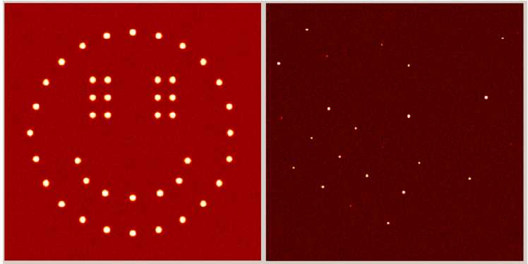
Figure 1. Two diffraction images - which is real?
Deliberate fabrication of crystallographic data has previously led to falsified structures being published and then later retracted from respected scientific journals1-3. Identified perpetrators, in these cases, had made very simple modifications to structural files, such as manually changing unit cell sizes and atom types, to produce adjusted data. Fortunately they were found to be unable to produce raw experimental data to support their claims. Kroon-Batenburg and Helliwell4 proposed that the requirement for the deposition of raw crystallographic data may be a potential method of preventing the submission of counterfeit structures. However we can show that the recreation of raw diffraction images is no longer difficult, opening the doors for those less scrupulous to take advantage, if this is not already occurring!
Detector frame formats from many manufacturers are well documented and this information can be reverse-engineered to encode synthetic diffraction data. This process was brought to light as a product of research into optimising data collection parameters for charge density studies. The chosen method required us to produce an algorithm which takes data from integrated .raw files as a starting point to create replicas of experimental images. A simple misuse of this code could take structure factors calculated for an entirely fabricated compound and produce diffraction images that, when processed, return the artificial structure. The frames are not visually distinguishable from authentic, experimentally determined, ones and can be fully integrated using standard protocols. The authors find this situation potentially alarming and requiring immediate attention.
A structure refined from data processed from these artificial diffraction images could pass all IUCr checkCIF5 protocols without raising alerts. We will present such a structure, full details of the algorithms employed and propose methodologies that may safeguard against this approach going undetected.
1. T. Liu et al. (2010). Acta Cryst. E66, e13-e14.
2. H. Zhong et al. (2010). Acta Cryst. E66, e11-e12.
3. International Union of Crystallography (2010). Acta Cryst. D66, 222.
4. L. M. Kroon-Batenburg & J. R. Helliwell (2014). Acta Cryst. D70, 2502-2509.
5. A. L. Spek (2009). Acta Cryst. D65, 148-155.
Keywords: Data, Simulation, Software,
12:35-13:00
Andreas Förster
EIGER HDF5 data and NeXus format
| Presentation (2.06 MB)
![[A. Foerster]](https://www.iucr.org/__data/assets/image/0017/113543/andreas_foerster.png)
Andreas Förster and Marcus Müller, Dectris Ltd, Neuenhoferstrasse 107, 5400 Baden, Switzerland
HDF5 is a container format designed for big data applications. In it, vast amounts of heterogeneous data can be stored in a small number of files that are easy to manage. Detectors of the EIGER series write datasets thousands of images big to HDF5 files and record most of the metadata that are required for data processing. The metadata are saved in a master file that is separate from the data but links to it. In this talk, I will present the HDF5 format and some of the metadata as written by EIGER detectors. I will also discuss metadata that are essential for processing but unknown to the detector and highlight blank fields that the EIGER HDF5 template provides for completion by beamline routines. A related talk by Herbert J. Bernstein [1] will explore ways of recording the geometry of the experimental setup. Software development, data processing, and effective archiving will all benefit from strict adherence to standards set by the NeXus committee.
[1] H. J. Bernstein "Metadata for characterising diffraction images: imgCIF and NeXus" in Workshop on Metadata for raw data from X-ray diffraction and other structural techniques, 22 - 23 Aug 2015, Rovinj, Croatia.
13:00-14:00
Lunch
Session III: Metadata for diffraction images and other experimental methods
14:00-14:25
Herbert J. Bernstein
Common diffraction image metadata specification in imgCIF, HDF5 and NeXus
| Presentation (1.09 MB)
![[H.J. Bernstein]](https://www.iucr.org/__data/assets/image/0020/113492/hjbernstein.jpg)
Herbert J. Bernstein
School of Chemistry and Materials Science, Rochester Institute of Technology, Rochester, NY, USA
The introduction of a new generation of fast pixel-array detectors, such as the Dectris Eiger and the Cornell-SLAC Pixel Array Detector (CSPAD), has required us to revisit and extend approaches we have used in the past to represent the data (the diffraction images) and the metadata (the information needed to reconstruct the experimental environment within which the data were collected) [1] [2]. For example, the axis descriptions from the imgCIF (image-supporting-CIF) dictionary have proven effective in reliably preserving the information about the frame-by-frame relative positions of beams, crystals and detectors and have been mapped into the context of HDF5 and NeXus to support the new Eiger format. See Andreas Förster's talk [3] for a discussion of the Dectris Eiger-specific HDF5/NeXus format. We are introducing a new, extended templating scheme to allow each beam line to specify the unique characteristics that will allow the metadata for a beam-line to be specified as either an HDF5/NeXus file or as an equivalent CBF/imgCIF file from which a site-file to be merged with run-specific data and metadata will be generated. A central repository of site-templates will be offered for convenience. This approach will help both in ensuring ease of processing of original data and in facilitating reliable handling of archived data.
[1] H. J. Bernstein, J. M. Sloan, G. Winter, T. S. Richter, NIAC, COMCIFS, "Coping with BIG DATA image formats: integration of CBF, NeXus and HDF5", Computational Crystallography Newsletter, 2014, 5, 12-18.
[2] A. S. Brewster, J. Hattne, J. M. Parkhurst, D. G. Waterman, H. J. Bernstein, G. Winter, N. K. Sauter, "XFEL Detectors and ImageCIF", Computational Crystallography Newsletter, 2014, 5, 19-25.
[3] A. Förster, M. Mueller, "EIGER HDF5 data and NeXus format", in Workshop on Metadata for raw data from X-ray diffraction and other structural techniques, 22-23 Aug 2015, Rovinj, Croatia.
Work supported in part by Dectris and by NIGMS.
14:25-14:50
Andrew Götz
Towards a generalised approach for defining, organising and storing metadata from all experiments at the ESRF
| Presentation (3.31 MB)
![[A. Goetz]](https://www.iucr.org/__data/assets/image/0018/113751/andy_gotz.jpg) Andrew Götz
Andrew Götz
European Synchrotron Radiation Facility, 71 Avenue des Martyrs, 38000 Grenoble, France
After more than 20 years of operation the situation concerning metadata at the ESRF is still very disparate between beamlines. The way the metadata required to analyse the raw data are defined and collected depends largely on the beamline concerned. Approaches vary from fully automated solutions implemented on the MX beamlines to a combination of automated and manual collection of metadata. This talk will not present the solution for MX (see talk by Gordon Leonard for more info) but will present a new approach for automating the collection and storing of well defined metadata for all experiments. The solution is based on a generic tool built at the ESRF which uses HDF5 for file format, Nexus for definitions (where possible) and icat for the metadata catalogue. The talk will present concrete examples of its use for nano tomography and fluorescence and radiation therapy. Ongoing work on how this will be extended to small angle scattering, coherent diffraction and eventually all other techniques will be presented. The talk will conclude with a discussion on the role of metadata in data policy and management.
14:50-15:15
John Westbrook
The PDB and experimental data
Presentation (7.45 MB)
15:15-15:35
Tea
15:35-16:00
Tom Terwilliger
Realising the Living PDB and how raw diffraction data and its metadata can help
| Presentation (57 kB)
![[T.C. Terwilliger]](https://www.iucr.org/__data/assets/image/0015/113721/terwilliger.jpg)
Thomas C. Terwilliger1 and Gerard Bricogne2
1Los Alamos National Laboratory, Mailstop M888, Los Alamos, NM 87545, USA. Email: terwilliger@lanl.gov
2Global Phasing Ltd, Sheraton House, Castle Park, Cambridge, CB3 0AX, UK. Email: gb10@globalphasing.com
The Protein Data Bank (PDB) is the definitive repository of macromolecular structural information. The availability of structure factors for most entries in the PDB has made it possible to continuously improve the models in the PDB by reinterpreting the primary data for existing structures as new methods of analysis, new biological information, and new ways of describing structures become available. This continuous improvement will be even more powerful once the diffraction images associated with each entry become accessible.
The key factor is that depositing raw images will stimulate the improvement of integration and processing software in the same way as the deposition of merged X-ray data hugely stimulated progress in refinement software. Revisiting deposited images with that improved software will deliver more accurate data (especially, free from the currently inadequate treatment of contamination by multiple lattices) against which to re-refine the deposited structures themselves.
With the initial interpretation of a structure, the original structure factors and the raw images, it will become possible both to carry out extensive validation of structures and to apply new algorithms for structure determination and analysis as they become available, leading to structures of ever-increasing accuracy and completeness.
Keywords: Structure quality; validation; PDB; automation; structure determination; raw data deposition
16:00
Close
Sunday 23 August
09:00
Open
Session IV: Data in the Wider World - From Laboratory to Database
09:00-09:25
Simon J. Coles
Diffraction Data in Context: metadata approaches
| Presentation (3.62 MB)
![[S.J. Coles]](https://www.iucr.org/__data/assets/image/0005/113549/Simon_Coles.JPG)
Simon Coles
Associate Professor and Director, UK National Crystallography Service, Chemistry, Faculty of Natural and Environmental Sciences, University of Southampton, Southampton, SO17 1BJ, UK
Diffraction experiments and the results arising from them must often sit in a given scientific context - e.g. in chemical crystallography, they are often performed as part of a study concerned with synthesising and characterising new compounds. The context for an experiment, i.e. why it has been performed, is often lost - particularly in the case where data is published on its own.
I will present approaches not only to ascribing metadata to the results of crystallographic experiments, but also to the general chemistry leading up to them. The first stages of work to build a model to support this have been published - http://www.jcheminf.com/content/5/1/52. I will go on to discuss recent work in two projects: (1) a collaboration with five big pharma companies, instrument manufacturers, electronic lab notebook vendors and the Royal Society of Chemistry to derive metadata for capturing the 'process' of performing experiments; and (2) a project (https://blog.soton.ac.uk/cream/) aimed at using metadata actively in the process of performing research, as opposed to purely for archival purposes. I will conclude with insights as to how the approaches taken in assigning metadata in these projects are important to consider when archiving and disseminating raw crystallographic data.
09:25-09:50
Suzanna Ward
CCDC metadata initiatives
| Presentation (4.15 MB)
![[S. Ward]](https://www.iucr.org/__data/assets/image/0020/113555/ward.jpg)
Suzanna Ward, Ian J. Bruno, Colin R. Groom and Matthew Lightfoot
Cambridge Crystallographic Data Centre, 12 Union Road, Cambridge, CB2 1EZ
For half a century the Cambridge Crystallographic Data Centre (CCDC) has produced the Cambridge Structural Database (CSD) to allow scientists worldwide to share, search and reuse small molecule crystal structure data. An entry in the CSD is often seen as 'just' a set of coordinates, but the associated metadata (data that describes and gives information about other data), is essential to contextualise an entry. Data that describes the substance studied, the experiment performed and the dataset as a whole are all vital.
This presentation, timed to coincide with the 50th anniversary of the CSD, will look at how metadata is used from deposition to dissemination of the CSD. We will look at how recent developments surrounding metadata have been targeted to improve the discoverability, validation and reuse of crystal structure data before looking to see what the future may hold.
09:50-10:15
Brian Matthews
Supporting Data Management Workflows at STFC
| Presentation (2.66 MB)
![[B. Matthews]](https://www.iucr.org/__data/assets/image/0010/113797/Matthews-Brian-09EC1353.jpg)
Brian Matthews, Scientific Computing, Rutherford Appleton Laboratory, Science and Technology Facilities Council, Harwell Oxford, Didcot OX11 0QX, UK
STFC has developed a systematic approach for managing and archiving data generated from its large-scale analytic facilities, which is used with variations by the ISIS Neutron Source, the Diamond Light Source and the Central Laser Facility. This is centred around the ICAT experiment metadata catalogue. The ICAT acts as a core middleware component recording and guiding the storage of raw data and subsequent access and reuse of the data; it has evolved into a suite of tools which can be used to build data management infrastructure. In this talk, I shall describe the current status of the ICAT.
The data rates and volumes generated from facilities are ever increasing and experimental science is becoming more complex. This is presenting challenges to the user community in accessing, handling and processing data. I shall describe some approaches to these problems and consider how we are exploring further support for data analysis and publication workflows within a large-scale facility. Finally, I shall consider how we might develop metadata to capture and share this information across communities.
10:15-10:40
Pierre Aller
Overview of metadata and raw data cataloguing at Diamond
| Presentation (1.48 MB)
![[P. Aller]](https://www.iucr.org/__data/assets/image/0007/113875/IYDdlsMXPierreAller02.JPG)
Pierre Aller and Alun Ashton, Diamond Light Source, Division of Science, Didcot, Oxfordshire, UK
Diamond Light Source as a relatively new facility has been able to capture and catalogue all its raw data (now over 3.6 Petabytes). Additionally as much metadata and processed data as possible has always been collated with the raw data and captured into both databases (ISPyB) for query and quick access, and into the raw data files (imgCIF/CBF and NeXus). Progress and status on these developments will be presented.
10:40-11:05
Brian McMahon
CODATA and (meta)data characterisation in the wider world
| Presentation (2.74 MB)
![[B. McMahon]](https://www.iucr.org/__data/assets/image/0004/113737/bm.jpg)
Brian McMahon
IUCr, 5 Abbey Square, Chester CH1 2HU, UK. Email: bm@iucr.org
This Workshop concentrates on scientific metadata and their importance in maximising the utility, trustworthiness and reuse of scientific data, especially to open doors to further avenues of study, and even new scientific insight. In a more general context, 'metadata' is a vehicle for categorising, classifying and collecting data sets. This presentation will review some of the organisations that take an interest in generic metadata and interoperability between metadata specifications from different disciplines or communities. The CODATA/VAMAS Working Group on Description of Nanomaterials provides a good example of collating different specialist metadata elements in a broad interdisciplinary framework. There will be a brief discussion of the granularity mismatch between generic and specialised metadata systems.
11:05-11:25
Coffee
Session V: What new metadata items are needed?
11:25-11:50
Gordon Leonard
What metadata is needed to make ESRF raw MX diffraction data intelligible for new users?
| Presentation (5.65 MB)
![[G. Leonard]](https://www.iucr.org/__data/assets/image/0016/113506/GLeonard.jpg)
Gordon Leonard
Structural Biology Group, European Synchrotron Radiation Facility, CS40220, 38043 Grenoble Cedex 9, France
The volume of diffraction data that can be collected during an experimental session on modern synchrotron-based Macromolecular Crystallography (MX) beamlines equipped with fast readout photon-counting pixel detectors and the rate at which it can be collected means it is currently difficult (or impossible) for users to manually process, during the experiment, all data sets collected. To help remedy this situation and to provide the at-beamline feedback that is sometimes necessary for a successful experiment 'autoprocessing' software[1,2] is often deployed with the results of automatic integration, scaling merging and reduction for individual data sets displayed in Laboratory Information Management Systems (LIMS) such as ISPyB[3] from where they can also be downloaded.
While the 'autoprocessing' approach works (i.e. provides data of sufficient quality for structure solution and refinement) in the vast majority of cases there is an increasing need for the post-experiment processing of raw diffraction images. In such cases the correct metadata for each dataset are essential to ensure the best results. For MX diffraction data collected at the ESRF this is stored in ISPyB, in the headers of the raw data images themselves and in automatically generated input files for the two main packages - XDS and MOSFLM - routinely used in the processing of ESRF-collected MX diffraction data. During my talk I will review the metadata currently logged during MX experiments at ESRF and look forward to what further metadata might be required when, either for validation purposes or the testing of new data processing and analysis protocols, raw data images are routinely made available to the wider scientific community.
[1] G. Winter et al. (2013). Acta Cryst. D69, 1260-1273. doi: 10.1107/S0907444913015308
[2] S. Monaco et al. (2013). J. Appl. Cryst. 46, 804-810. doi:10.1107/S0021889813006195
[3] S. Delageniere et al. (2011). Bioinformatics, 27, 3186-3192. doi:10.1093/bioinformatics/btr535.
11:50-12:15
Matthew Blakeley
What metadata is needed to make Institut Laue Langevin neutron diffraction raw data intelligible for new users?
| Presentation (8.15 MB)
![[M. Blakely]](https://www.iucr.org/__data/assets/image/0005/113765/blakely.jpg)
Matthew Blakeley
Institut Laue-Langevin, 71 Avenue des Martyrs, 38000 Grenoble, France
Central facilities for neutron scattering and synchrotron X-ray sources in Europe are working together to develop and share infrastructure for the data collected there. Such co-operation should make it easier and more efficient for users to access and process their data, and provide more secure means of storage and retrieval. It should also increase the scientific value of the data by opening it up to a wider community for further analysis and fostering new collaborations between scientific groups. However, with these developments comes a need to define how raw data are stored and made accessible, and in particular, what metadata are included to allow diffraction data to be intelligible to new users. To this end, an ILL data policy (https://www.ill.eu/fr/users/ill-data-policy/) was established in 2012, and a number of tools (e.g. [i] https://data.ill.eu [ii] https://logs.ill.eu) are being developed. These currently allow experimental data (identified by a DOI) to be consulted and downloaded remotely and ultimately will allow for (re)processing and validation of experimental data.
12:15-12:40
Kamil Dziubek
Metadata in high-pressure crystallography
| Presentation (1.13 MB)
![[K. Dziubek]](https://www.iucr.org/__data/assets/image/0015/113505/dziubek.jpg)
Kamil F. Dziubeka and Andrzej Katrusiakb
aLENS - European Laboratory for Non-Linear Spectroscopy, Sesto Fiorentino, Italy
bFaculty of Chemistry, Adam Mickiewicz University, Poznań, Poland
The deposition of metadata related to specific techniques used for crystallographic experiments can be simplified by formulating guidelines for their preparation. One of the experimental techniques quickly gaining ground in the field of crystallographic research is high-pressure diffraction studies. They involve additional equipment for pressure generation, pressure calibration, etc. The high pressure cell can interfere with the primary or diffracted beam, which can contaminate the diffraction patterns and introduce errors in reflection intensities. The experimental details are vital for the evaluation and analysis of the data, and therefore the metadata are needed to be stored along with the raw diffraction images.
The most essential descriptors concern: (1) orientation of the pressure cell with respect to the incident beam and the detector; (2) the sample preparation and shape, both for powder and single crystals; (3) a reference to the high-pressure vessel, and for unique equipment the dimensions of its relevant components, such as the anvil design, gasket thickness, chamber diameter, backing-plate type; (4) chemical composition of the cell parts, e.g. anvils, gasket and backing plates, pressure-transmitting medium; (5) the method of fixing the sample in the high-pressure chamber, if used; (6) the method of positioning the pressure cell during the data acquisition; (7) the pressure-measurement method. This information is indispensable for reproducing the results of structural refinements from the raw data or for attempting other methods of refinement. The pressure transmitting medium can dramatically change the sample compression, due to its possible interaction with the sample (such as penetration into the pores) or hydrostatic limit of the medium. The sample history can also affect the results. If the sample was recrystallized in situ in isothermal or isochoric conditions, from solution or melt, the details of the crystallization protocol should be provided. Simple edition rules and a checklist can considerably simplify the deposition of metadata and increase their informative value.
The authors are representing the IUCr Commission on High Pressure, AK is the chair of the Commission. KFD gratefully acknowledges the Polish Ministry of Science and Higher Education for financial support through the "Mobilność Plus" program.
12:40-13:40
Lunch
Session VI: Metadata schemas
13:40-14:05
James Hester
Creating and manipulating universal metadata definitions
| Presentation (1.59 MB)
![[J.R. Hester]](https://www.iucr.org/__data/assets/image/0004/113692/James_Hester.jpg) James R. Hester
James R. Hester
Australian Nuclear Science and Technology Organisation, New Illawarra Road, Lucas Heights, NSW 2234, Australia
Metadata discussions are often closely linked to particular formats. However, facts about the natural world cannot depend on the medium used to transmit those facts. It follows that we are able to completely describe our metadata without reliance on any particular file format, and that we can distil metadata definitions from pre-existing data transfer frameworks regardless of the particular format used. This promising, if obvious, general conclusion does not specify what information needs to be provided in our format-free metadata definition. Following Spivak and Kent (2012) I suggest that it is sufficient that the metadata definitions can be expressed as functions mapping some domain to some range.
This talk will explore some of the implications of this approach, including the independence of file format and metadata specification, specification of algorithms for interconversion between data files in differing formats, unification of disparate metadata projects, and simple steps to produce a complete metadata description.
Spivak D.I., Kent R.E. (2012) "Ologs: A Categorical Framework for Knowledge Representation." PLoS ONE, 7(1):e24274. doi:10.1371/journal.pone.0024274
14:05-14:30
Brian McMahon
The Crystallographic Information Framework as a metadata library
| Presentation (2.50 MB)
Brian McMahon
IUCr, 5 Abbey Square, Chester CH1 2HU, UK. Email: bm@iucr.org
The Crystallographic Information File (CIF) was introduced as a data exchange standard in crystallography in 1991 [1] and has become embedded in the practice of single-crystal and powder diffractometry. Versions of CIF exist that describe macromolecular structure and diffraction images [2], so that CIF may be used anywhere in a data pipeline from image capture to publication in what has been called a 'coherent information flow' (also CIF!) [3].
In practice, slight differences in data model and file format have led to 'dialects' of CIF, which can coexist quite happily. However, there is a consequent barrier to full interoperability. A new version of the CIF format [3] will allow the development of a new generation of CIF 'dictionaries' (the formal data description schemas or 'ontologies'). This will allow fully automatic interconversion between existing CIF data files in either formalism, but has the added bonus of providing a descriptive framework for any type of crystallographic information. Formally, the CIF approach makes no distinction between 'data' and 'metadata', and so is arbitrarily adaptable and extensible to any domain of structural science or further afield. Since the CIF format has a very simple syntactic structure which makes the contents very easy to read, CIF dictionaries can provide a simple template for developing new metadata schemas by working scientists who are not experts in informatics.
[1] Hall, S. R., Allen, F. H. & Brown, I. D. (1991). The Crystallographic Information File (CIF): a New Standard Archive File for Crystallography. Acta Cryst. A47, 655-685.
[2] Hall, S. R. & McMahon B. (eds) (2005). International Tables for Crystallography, Volume G: Definition and exchange of crystallographic data. Dordrecht: Springer. Corrected reprint (2010). Chichester: Wiley.
[3] McMahon, B. (2013). A coherent information flow in crystallography. Presentation at ECM28 Satellite Symposium on Crystallographic Information and Data Management, U. Warwick, UK. See also https://youtu.be/BiYETNUbfVo
14:30-14:40
General discussion
14:40-15:00
Tea
15:00-15:50
Practical session: building a metadata description
Presentation 1 (1.48 MB) | Presentation 2 (2.87 MB)
16:00
Close
18:00
ECM29 Opening Ceremony